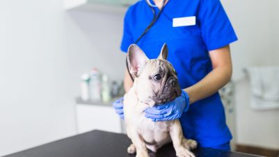
Recognising, diagnosing and treating ureteral obstruction presents a challenge. In recent years, the subcutaneous ureteral bypass (“SUB” device) (Figure 1) has become the treatment of choice for many veterinary surgeons, particularly for cases of ureteral obstruction in cats. However, this procedure is not without its complications and drawbacks. This begs the question – is there a better alternative?
Recognition
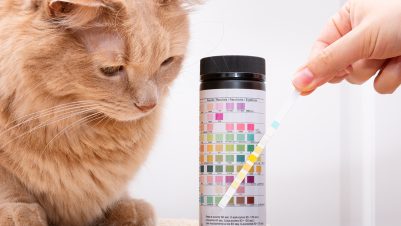
Recognising and diagnosing ureteral obstruction can be difficult as the history is often non-specific, and animals can present with vague clinical signs. The severity of clinical signs may also depend on the degree of function of the contralateral kidney and the animal’s ability to compensate for unilateral obstruction. Frequently, signs include lethargy, anorexia, vomiting, polyuria and polydipsia. Abdominal pain is common, particularly on renal palpation.
Animals may have a palpably enlarged kidney (or kidneys if there is acute bilateral obstruction), but this may not always be the case, especially if there is chronic renal fibrosis. Palpation of “big-kidney-little-kidney” can raise suspicion of a previous occult renal injury unilaterally, with illness only becoming evident when the remaining kidney is challenged.
While it is important to remember that acute-on-chronic kidney disease is a possibility […] ureteral obstruction should be included as a differential diagnosis in any case presenting with acute azotaemia or a rapid clinical deterioration
Azotaemia is almost always present on biochemical analysis and there are often electrolyte abnormalities, including hyperkalaemia and hyperphosphataemia. Cases of ureteral obstruction may be confused with the onset or progression of chronic kidney disease in older animals. While it is important to remember that acute-on-chronic kidney disease is a possibility in these animals (among the myriad of possible conditions, including bacterial pyelonephritis and glomerulonephropathies), ureteral obstruction should be included as a differential diagnosis in any case presenting with acute azotaemia or a rapid clinical deterioration, especially for small-breed dogs and cats.
Diagnostic imaging
Diagnostic imaging for this condition generally relies on radiography and/or ultrasonography but bear in mind that the sensitivity of ultrasonography to detect ureteral obstruction is operator dependent. Occasionally, one or multiple ureteroliths may be evident on plain abdominal radiographs. The absence of a visible ureterolith does not rule out ureteral obstruction. This is because they can be small (the internal diameter of the feline ureter measures approximately 0.4mm), and ureteral strictures are not detectable radiographically (strictures are a feature of obstruction in over 25 percent of cats). Secondary changes such as renomegaly may be recognised.
In a referral setting, diagnosis is commonly made based on consistent serum biochemistry and specialist ultrasonography or using three-dimensional imaging
Hydronephrosis, pyelectasia or hydroureter are generally apparent via ultrasonography. Skilled sonographers may identify ureteroliths or note a narrowing of the ureters consistent with a stricture. It is important to note that not all ureteral obstructions lead to marked pyelectasia or renomegaly, particularly in cats with pre-existing chronic kidney disease, as renal fibrosis can limit the expected expansion. In a referral setting, diagnosis is commonly made based on consistent serum biochemistry and specialist ultrasonography or using three-dimensional imaging (particularly computed tomography excretory urography (CT IVU)).
Treatment for ureteral obstruction
Animals with confirmed ureteral obstruction are all potential surgical candidates as medical management (Table 1) alone is rarely successful, resolving obstruction in only 8 to 13 percent of cases (Lulich et al., 2016; Kyles et al. 2005). Initial stabilisation is an important first step to manage hypovolaemia, dehydration, azotaemia and significant electrolyte imbalances. Serum creatinine level at presentation has recently been associated with survival times in one large study of cats undergoing SUB placement in the UK (Kulendra et al., 2021). The median survival time of cats with a serum creatinine of 440μmol/l or above (IRIS acute kidney injury (AKI) stage four and five) was 530 days, compared to a median survival time of 949 days for those cats presenting with creatinine levels under 440μmol/l (IRIS AKI stage one to three).
| Drug | Dosage |
|---|---|
| Intravenous fluid therapy | Appropriate rates, crystalloids – careful monitoring for fluid overload |
| Mannitol | 0.25g/kg bolus over 30 to 60 minutes followed by CRI at 1mg/kg/min for 24 hours – care must be taken in cats with cardiac disease |
| Prazosin | 0.25mg/cat BID, 0.5 to 2mg/dog BID – blood pressure should be monitored |
| Analgesia | Methadone at 0.2mg/kg (or buprenorphine at 0.01 to 0.02mg/kg in cats) |
| Antibiotics | Administered only if there is evidence of a urinary tract infection (33 percent of cats are reported to have positive urine cultures) |
Analgesia and management
Opioid analgesia is essential for ureteral obstruction as it is a highly painful condition. Isotonic crystalloid intravenous fluid therapy type and administration rate are guided by clinical assessment of volume status, hydration and urine production. Regular reassessment is necessary to avoid fluid overload and to assess treatment response. Diuresis should be encouraged, with the aim of passing the urolith by an increase in urine flow, using furosemide or mannitol. Note that mannitol may be contraindicated in anuric animals due to the risk of significant volume overload. Serum creatinine is serially monitored, and ureteral/renal ultrasound should be repeated.
If the obstruction cannot be relieved in 48 hours, surgery should be considered, as prolonged obstruction risks progressive renal injury. Of course, it is often not possible to know how long the obstruction was present before presentation. In the author’s experience, single stones or debris close to the ureterovesical junction (and if the contralateral kidney function is good) are more likely to resolve with medical management than cases of stricture, multiple uroliths or a defunct contralateral kidney.
If the obstruction cannot be relieved in 48 hours, surgery should be considered, as prolonged obstruction risks progressive renal injury
Careful management of owner expectations is absolutely essential prior to surgery, as complication rates for treating ureteral obstructions are high and persistent renal disease can cause ongoing morbidity and limit life expectancy. Particularly in the case of SUB, considerable post-operative care with regular veterinary rechecks to flush the device are required. Some animals will not tolerate this, and there are attendant travel/cost implications for owners.
Surgical options
Traditional surgical options for ureteral obstruction include ureterotomy (Figure 2), ureteronephrectomy, and ureteral resection (Figure 3) and reimplantation. SUB placement is advocated as the new treatment of choice due to the high reported complication rates for traditional surgeries. However, there is a moderately high perioperative complication rate. Some studies also report significant medium- to long-term complications, with only relatively short post-operative survival rates (medians in the region of one to two years).


In a recent UK study of cats undergoing SUB placement for ureteral obstructions, 10 percent did not survive to discharge, while 48 percent experienced “major” complications (Kulendra et al., 2021). These complications included infection or technical issues requiring revision surgery or death/euthanasia, with the majority of these occurring after discharge from hospital. The median survival time in this population was 820 days. An earlier 2020 study reported a median survival time of 274 days (with a range of 1 to 311) and an 80 percent complication rate (Vrijsen et al., 2021).
Careful case selection is essential, and owner expectations should be managed. In the author’s experience, many owners either elect euthanasia if medical management fails or sometimes pursue traditional surgical options if the features of the disease make this possible (eg a single unilateral ureterolith may be amenable to ureterotomy with or without an intraluminal stent). This decision is due to multiple factors, including:
- the condition of the patient at the time of ureteral obstruction diagnosis
- the risk of renal function not recovering post-operatively
- the cost of surgery
- the burden of future aftercare
The SUB
The SUB 3.0 device consists of two locking loop pigtail catheters connected to a port placed subcutaneously. One catheter is placed into the renal pelvis and the other into the bladder, allowing urine to bypass the diseased ureter and relieve the obstruction. Placement is most commonly guided by intraoperative fluoroscopy and, occasionally, via ultrasonography. Active urinary tract infections risk biofilm formation on the permanent device, further complicating aftercare. It is expected that veterinary surgeons receive specific training to place these devices to minimise perioperative complications.
Post-operative care
The SUB device should be flushed every three to six months for the rest of the patient’s life. While SUB devices are often placed in referral hospitals, owners may prefer to revisit their primary care practice for flushing. Some animals may tolerate conscious flushing, but sedation is commonly required. The port is palpated subcutaneously on the ipsilateral ventral abdomen, and a urine sample is obtained using aseptic technique to insert a Huber needle (a sterile non-coring needle) perpendicularly through the skin into the silicone insertion site in the centre of the port. A small volume of agitated sterile saline is then injected into the port while the renal pelvis is monitored with ultrasonography, ensuring the renal pelvis does not become over-distended. Once turbulence is seen in the renal pelvis, the fluid is withdrawn and the bladder is monitored on repeat flushing.
While SUB devices are often placed in referral hospitals, owners may prefer to revisit their primary care practice for flushing
Once patency has been confirmed, a solution of tetrasodium EDTA or taurolidine citrate is instilled. This aims to prevent the formation of a biofilm. Approximately 1 to 2ml should be injected slowly in pulses to allow it to drain the length of the tubes, ensuring there is no renal pelvis dilation. The urine sample should be cultured as early identification of any infection increases the likelihood of successful treatment.
Survival rates
Recently, reported survival times vary widely from 1 to 1,915 days (Kulendra et al., 2021; Vrijsen et al., 2021). Median survival times between 274 and 820 days are expected for cats (Kulendra et al., 2021; Vrijsen et al., 2021). Most complications of ureteral obstruction treatment occur after discharge, so careful owner monitoring and regular veterinary rechecks are essential. Initially, the SUB device is flushed at surgery and the following day, then after three days, one week and three weeks, and then every three months. This should reduce the risk of complications such as urinary tract infection, biofilm formation and tube obstruction (with clots or by mineralisation).
Summary
While SUB placement is a good option for cases of ureteral obstruction with multiple immobile uroliths and conditions affecting the entirety of the ureter or stricture, the complication rate is high, and commitment on the part of the owner and the veterinary surgeon is essential. Traditional surgical techniques remain an option for some animals as these require less commitment outside the immediate post-operative period. For example, when performed by a skilled surgeon, ureterotomy may still be the preferred option for a single urolith, and ureteral resection and reimplantation can treat a distal ureterolith or stricture.