| M0: Inactive Healthy skin, ensure interdigital area clear, no evidence of previous lesions |
 | M1: Active A small (less than 2cm) focal lesion. Erosive or early ulcerated stage |
 | M2: Active Acute and painful ulceration, may have grey exudate or granulomatous changes |
 | M3: Inactive Healing lesion, often seen after topical treatment, flat and benign in appearance. Can regress in favourable conditions or evolve towards chronic lesion |
 | M4: Chronic (Inactive) Chronic hyperkeratotic lesion which may have filamentous proliferations. Bacteria can be isolated from these lesions |
 | M4.1: Active Chronic hyperkeratotic lesion with re-emerging active ulcerative area |
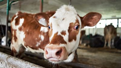
Digital dermatitis (DD) is a superficial, painful, contagious inflammation of the skin of the bovine foot mainly affecting the hind feet. It was first identified in Italy in 1974 and subsequently in the UK in 1987. It is now considered endemic in the UK and is considered to have a prevalence of 15 to 43 percent in infected herds (Solano et al., 2017). The impact of DD is significant as it can cause severe pain (Bruijnis et al., 2012) and reduced milk yields and reproductive performance (Bruijnis et al., 2010; Cha et al., 2010; Ettema et al., 2010; Evans et al., 2016).
The infection is multifactorial with varied species of bacteria isolated from active lesions. Treponeme species are commonly isolated with the three species most prevalent being Treponema medium, T. phagedenis and T. pedis. The treponemes use damage to the epidermis and dermis (often due to contact with slurry) to invade, often via hair follicles and sebaceous glands.
Lesions can be very variable in appearance with the most common being a circumscribed ulcerated or granulomatous area that can take the appearance of hairy warts due to hyperkeratosis. Lesions can be classified by the M-stage scoring system (Table 1; Dopfer et al., 1997; Berry et al., 2012).
New infections typically take many weeks to develop, with a recent study demonstrating a mean of 133 days, while reactivation of M4 lesions can be very rapid with re-ulceration seen within days (Krull et al., 2016).
Impact
Welfare
Lameness associated with DD (mobility score 2 or 3) is associated with active clinical lesions.
DD had the highest welfare impact of the foot disorders rated in a study by Bruijnis et al. (2012), and this research found that the impact of subclinical foot issues was nearly equivalent to that of clinical disease. A Swedish study assessed welfare with a statistically significant change in lying time (Pavlenko et al., 2011).
Economics
All lameness has an economic impact through both increased costs (expenditure) and reduced returns (losses) (Table 2). It is important that both clinical and subclinical disease is considered when looking at the economic impact of DD, as it is estimated that 32 percent of total costs due to foot disorders are due to subclinical disorders (Bruijnis et al., 2010) and this may be an area that is underestimated on UK farms.
| Expenditure | Losses |
|---|---|
| Treatment | Reduced milk production |
| Prevention | Reduced reproductive performance |
| Increased risk of culling and death | |
| Reduced welfare effects |
An impact on milk yield has also been demonstrated: Pavlenko et al. (2011) recorded an average reduction of 5.5kg of energy-corrected milk (ECM) production for up to three weeks post-diagnosis with a clinical DD lesion.
Another study examined the hind feet of all lactating cows from 47 French dairy farms monthly for six months, and found that milk yield was reduced in both primiparous and multiparous animals, with both moderate and severe disease (Table 3; Relun et al., 2013). The DD status of each animal was defined by three categories: no DD (scores of M0 and/or M4 on both feet), moderate (score of M1 on one or both feet) and severe (score of M2 on one or both feet).
| Primiparous | Multiparous | |
|---|---|---|
| Moderate DD | 0.63kg per day | 0.5kg per day |
| Severe DD | 0.5kg per day | 0.75 kg per day |
In heifers, DD lesions that occur before calving have an impact on the animal’s first lactation performance in terms of 305-day milk yield, calving to conception time and conception rate to first artificial insemination (AI) (Table 4; Gomez et al., 2015).
| DD in six months pre-calving | Days calving to conception | Conception rate to first AI | 305-day milk yield (kg) |
|---|---|---|---|
| Type I (no DD lesions) | 132 | 42.3 percent | 12,464 |
| Type II (one DD event) | 134 (+2) | 36.3 percent (-6 percent) | 12,265 (-199) |
| Type III (multiple DD events) | 157 (+25) | 29 percent (-13.3 percent) | 12,129 (-335) |
It has been demonstrated that M1 and M2 were 27 times more likely to remain as M2 lesions and significantly more likely to be M4 rather than M0 a month later (Berry et al., 2012). M4 cows were more likely to remain as M4 or become an M4.1. When the contribution of different M scores to transmission of DD was assessed, it was demonstrated that M4 is responsible for 88 percent of the R0 (Biemans et al., 2018).
Control
Control should focus on preventing infection to minimise active lesions that progress to chronic lesions which maintain the infection pressure, as well as prompt and effective treatment of clinical lesions.
Biosecurity, at both external and internal levels, is paramount. Ensure any bought-in animals are isolated, with active lesions treated, and that they are foot bathed before entry into the herd.
Control should focus on preventing infection to minimise active lesions that progress to chronic lesions which maintain the infection pressure, as well as prompt and effective treatment of clinical lesions
Foot-trimming equipment is a recognised reservoir of bacteria, and disinfection is therefore important. Ensure knives are disinfected between animals and even between feet of an individual. Disinfection requires a minimum of 20 seconds contact time. Disinfectants that are effective include 1:100 FAM30, 2 percent Virkon or 2 percent sodium hypochlorite (Gillespie et al., 2020).
Foot bathing is commonly used both to disinfect the feet to reduce infection pressure, and to treat lesions. Foot baths need to allow smooth cow flow with good contact time and coverage of claws and interdigital skin. It is important that the biocide is added at the correct concentration and replenished regularly. The choice of biocide is beyond the scope of this article, but it is important to note that the off-label use of antibiotics is no longer justifiable.
Treatment
Whole herd treatment
A review of 13 studies could not support an association between herd treatment and a beneficial effect in the prevention and treatment of DD (Ariza et al., 2017).
However, a more recent UK study examined “blitz” treatment in which all cows with lesions were treated with concurrent and subsequent improvements in hygiene and efficacy of foot bathing (Pedersen, 2019). Initially, 26 percent of cows had an active or recurring lesion; this reduced to 2 percent at six weeks, and further reduced to 0.5 percent by five months. By the 14-month review it remained significantly lower than the original figure, at 6 percent.
Individual animal treatment
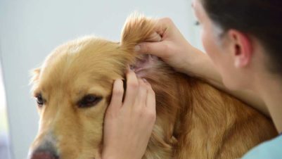
Identification is key and, if done in the parlour, this requires a good light source as well as a mirror to examine the interdigital skin. As 80 to 90 percent of lesions are seen in the hind feet, assessment in the parlour can be justified.
DD lesions should be cleaned, dried and debrided before the application of a topical antibacterial preparation. If debulking of proliferative tissue is required, local anaesthetic should be used (Bell and Vanhoudt, 2020). The choice of topical treatment is once more beyond the scope of this article.
Pain management
In contrast to claw horn lesions where the benefit of NSAIDs has been demonstrated (Thomas et al., 2015), until recently there was little data to support the use of NSAIDs in DD, despite the evidence that it is painful and impacts on welfare and production.
Until recently there was little data to support the use of NSAIDs in DD, despite the evidence that it is painful and impacts on welfare and production
Kasiora et al. (2021) evaluated the use of ketoprofen for the treatment of DD in dairy cattle. This was a randomised positive-controlled clinical trial. Following a mobility score (MS), cows with a DD lesion were enrolled. All lesions were cleaned and dried, and an oxytetracycline spray topically applied. Treated animals (T) had a single intramuscular injection of ketoprofen, while control animals (C) did not. The MS at enrolment and at a week post-intervention were recorded, as well as the daily milk yield for a week before and week after.
When only animals that were lame at enrolment were considered, C cows were 20 times more likely than T cows to be lame a week later. Overall, T cows produced 2.98kg more milk than C cows. There was a numerical increase of 5kg of milk for lame T compared to lame C cows in the early and late lactation group. When only lame fresh (one week post-calving) cows were considered, there was a 10.49kg increase in milk yield (T = 58.38, C = 47.89, p<0.05).
Conclusions
DD is an endemic and painful cause of lameness that impacts on welfare and production of dairy cattle. Excellent biosecurity with scrupulous hygiene is key to minimising disease. Treatment must be prompt and effective, after examination of the whole foot. Recent UK work supports the use of ketoprofen to manage the associated pain and inflammation.